Beranda
/ Rib Cage Anatomy - Superficial And Deep Muscles Of The Shoulder And Rib Cage Medical Stock Images Company : Rib cage pain may be sharp, dull, or achy and felt at or below the chest or above the navel on either side.
Rib Cage Anatomy - Superficial And Deep Muscles Of The Shoulder And Rib Cage Medical Stock Images Company : Rib cage pain may be sharp, dull, or achy and felt at or below the chest or above the navel on either side.
Insurance Gas/Electricity Loans Mortgage Attorney Lawyer Donate Conference Call Degree Credit Treatment Software Classes Recovery Trading Rehab Hosting Transfer Cord Blood Claim compensation mesothelioma mesothelioma attorney Houston car accident lawyer moreno valley can you sue a doctor for wrong diagnosis doctorate in security top online doctoral programs in business educational leadership doctoral programs online car accident doctor atlanta car accident doctor atlanta accident attorney rancho Cucamonga truck accident attorney san Antonio ONLINE BUSINESS DEGREE PROGRAMS ACCREDITED online accredited psychology degree masters degree in human resources online public administration masters degree online bitcoin merchant account bitcoin merchant services compare car insurance auto insurance troy mi seo explanation digital marketing degree floridaseo company fitness showrooms stamfordct how to work more efficiently seowordpress tips meaning of seo what is an seo what does an seo do what seo stands for best seotips google seo advice seo steps, The secure cloud-based platform for smart service delivery. Safelink is used by legal, professional and financial services to protect sensitive information, accelerate business processes and increase productivity. Use Safelink to collaborate securely with clients, colleagues and external parties. Safelink has a menu of workspace types with advanced features for dispute resolution, running deals and customised client portal creation. All data is encrypted (at rest and in transit and you retain your own encryption keys. Our titan security framework ensures your data is secure and you even have the option to choose your own data location from Channel Islands, London (UK), Dublin (EU), Australia.
Rib Cage Anatomy - Superficial And Deep Muscles Of The Shoulder And Rib Cage Medical Stock Images Company : Rib cage pain may be sharp, dull, or achy and felt at or below the chest or above the navel on either side.. Humans usually have 24 ribs, in 12 pairs. The remainder of the rib is the body of the rib (shaft). The rib cage is more like an egg because the top is narrower than the bottom. Rib cage anatomy watercolor print, rib cage anatomy watercolor art, chest bones anatomy, thorax anatomy art print as85 genefyart. See more ideas about anatomy reference, anatomy drawing, human anatomy.
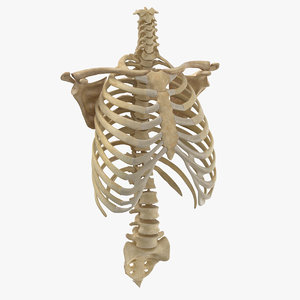
The rib cage is the arrangement of ribs attached to the vertebral column and sternum in the thorax of most vertebrates, that encloses and protects the vital organs such as the heart, lungs and great vessels. In the anatomical position, the angles align with the medial border of the scapula. As viewed from the side, the thoracic spine's vertebrae form a kyphotic curve that runs from t1 to t12, in which the spine curves outward towards the back of the body to allow more room for the internal organs such. Instead, anatomists classify the ribs as flat bones, and they are located within the axial skeleton. There are twelve (12) pairs of ribs and all articulate posteriorly with the thoracic vertebrae.
3d Rib Cage Models Turbosquid from static.turbosquid.com The ribs are long, flat, curved bones that form most of the thoracic cage. The rib below that is rib 2, and it connects to the t2 thoracic vertebra, and so on. At the chest, many rib bones connect to the sternum via costal cartilage,. Instead, anatomists classify the ribs as flat bones, and they are located within the axial skeleton. From the anatomy of the human rib cage, we can tell that the human ribs bones have several parts: Introduction to the structure of the ribcage and ribs: The ribs also provide attachment sites for thoracic muscles. It is not unusual for people with trisomy 21 (down syndrome) , for example, to have an extra or a missing pair of ribs, and the rib abnormalities in these cases rarely.
Just lateral to the tubercle is the angle of the rib, the point at which the rib has its greatest degree of curvature.
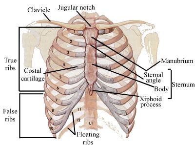
They articulate with the vertebral column posteriorly, and terminate anteriorly as cartilage (known as costal cartilage). Ten of the twelve ribs connect to strips of hyaline cartilage on the anterior side of the body. The remainder of the rib is the body of the rib (shaft). As part of the bony thorax, the ribs protect the internal thoracic organs. The rib cage is more like an egg because the top is narrower than the bottom. The head of the rib includes the articular surface, facies articularis capitis costae. The ribs are a set of twelve paired bones which form the protective 'cage' of the thorax. There are twelve pairs of ribs, all of which articulate with the vertebral column, while only the first seven ribs directly articulate with the sternum. Head (caput costae) neck (collum costae) body, corpus costae; The cervicothoracic junction is where the neck (cervical spine) connects with the upper back (thoracic spine). And the front part of the rib. Introduction to the structure of the ribcage and ribs: Abdominal anatomy organs in quadrants.
And the front part of the rib. 5 out of 5 stars (81) $ 16.71. The rib below that is rib 2, and it connects to the t2 thoracic vertebra, and so on. At the chest, many rib bones connect to the sternum via costal cartilage,. They articulate with the vertebral column posteriorly, and terminate anteriorly as cartilage (known as costal cartilage).
Rib Cage Medical Art Library from medicalartlibrary.com Humans usually have 24 ribs, in 12 pairs. Ten of the twelve ribs connect to strips of hyaline cartilage on the anterior side of the body. In this video we discuss the structure of the rib cage or thoracic cage. The cartilage strips are called costal cartilage (costal is the anatomical adjective that refers to the rib) and connect on their other end to the sternum. In the anatomical position, the angles align with the medial border of the scapula. Rib cage pain may be sharp, dull, or achy and felt at or below the chest or above the navel on either side. But for an anatomy study, it's not. The ribs also provide attachment sites for thoracic muscles.
Rib deformities may appear in isolation, with no other anatomical anomalies, or in association with other problems, sometimes as part of an identified condition or syndrome.
Anatomy of human stomach 10 photos of the anatomy of human stomach anatomy human colon, anatomy human digestive system, anatomy human heart, anatomy human kidney, anatomy human liver, anatomy human pancreas, anatomy human spleen, human body stomach, stomach, anatomy human colon, anatomy human digestive system, anatomy. As part of the bony thorax, the ribs protect the internal thoracic organs. Anatomy, rib cage, my heart is a flower, rib cage art, medical art, gift for doctor, botany, anatomical art, book page art, book art ambicerebral. The cervicothoracic junction is where the neck (cervical spine) connects with the upper back (thoracic spine). See more ideas about anatomy reference, anatomy drawing, human anatomy. As viewed from the side, the thoracic spine's vertebrae form a kyphotic curve that runs from t1 to t12, in which the spine curves outward towards the back of the body to allow more room for the internal organs such. Head (caput costae) neck (collum costae) body, corpus costae; Related posts of rib cage diagram with organs anatomy of human stomach. There are twelve (12) pairs of ribs and all articulate posteriorly with the thoracic vertebrae. Ten of the twelve ribs connect to strips of hyaline cartilage on the anterior side of the body. From the anatomy of the human rib cage, we can tell that the human ribs bones have several parts: The head of the rib includes the articular surface, facies articularis capitis costae. Rib deformities may appear in isolation, with no other anatomical anomalies, or in association with other problems, sometimes as part of an identified condition or syndrome.
Ten of the twelve ribs connect to strips of hyaline cartilage on the anterior side of the body. It may occur after an obvious injury or without explanation. In this video we discuss the structure of the rib cage or thoracic cage. Introduction to the structure of the ribcage and ribs: They articulate with the vertebral column posteriorly, and terminate anteriorly as cartilage (known as costal cartilage).
Skeletal Series Part 5 The Human Rib Cage These Bones Of Mine from thesebonesofmine.files.wordpress.com Rib cage pain may be sharp, dull, or achy and felt at or below the chest or above the navel on either side. The first rib is attached to thoracic vertebra. Rib cage pain can be caused. It is made up of 12 pairs of ribs. The ribs are a set of twelve paired bones which form the protective 'cage' of the thorax. The ribs can be classified based on their articulations: It has clear front, side, and back planes. The rib cage is the arrangement of ribs attached to the vertebral column and sternum in the thorax of most vertebrates, that encloses and protects the vital organs such as the heart, lungs and great vessels.
The ribs also provide attachment sites for thoracic muscles.
In this video we discuss the structure of the rib cage or thoracic cage. It is not unusual for people with trisomy 21 (down syndrome) , for example, to have an extra or a missing pair of ribs, and the rib abnormalities in these cases rarely. Rib cage pain may be sharp, dull, or achy and felt at or below the chest or above the navel on either side. The angles of the ribs form the most posterior extent of the thoracic cage. Rib deformities may appear in isolation, with no other anatomical anomalies, or in association with other problems, sometimes as part of an identified condition or syndrome. Rib cage anatomy watercolor print, rib cage anatomy watercolor art, chest bones anatomy, thorax anatomy art print as85 genefyart. The ribs also provide attachment sites for thoracic muscles. From the anatomy of the human rib cage, we can tell that the human ribs bones have several parts: Humans usually have 24 ribs, in 12 pairs. The cervicothoracic junction is where the neck (cervical spine) connects with the upper back (thoracic spine). See more ideas about anatomy reference, anatomy drawing, human anatomy. But for an anatomy study, it's not. In the anatomical position, the angles align with the medial border of the scapula.